SPONDYLOS menu →


Osteoporotic Kyphosis. What is it?

Osteoporosis is a condition that affects about 1 in 10 postmenopausal women.
Probably due as in almost all conditions in gene. There are of course several factors that enhance the development of the condition, such as problems of the thyroid gland, kidney problems, tranquilizers, failure to exercise, not eating dairy, pregnancies, breast-feeding, failure to exercise, use of cortisone and many others .
The correct information, the early diagnosis and the proposed treatment to control it, for not cause fractures of the vertebrae and osteoporotic kyphosis.

In Osteoporosis we have a progressive reduction of bone resistance, especially cancellous bone, with a reduction in bone trabeculae and a “vacating” of the bone.
In the vertebrae bodies, which are predominantly cancellous bone, this condition is definitive for the static ability of the spine.
The reduction of the bone trabeculae and the resulting increase of hollows weaken the vertebra bodies, which begin to form microscopic fractures (caving).
This causes sudden intense pain, and the body cannot bear the pressures of the body weight.

The continuation of axial compression as well as the anterior torques of bending forces that are exerted on the vertebrae bodies leads to a sphenoid deformation. If no action is taken, as far as the increase of the compression is concerned, this phenomenon is continued in another vertebra, resulting in continuously greater kyphosis and entering in a vicious circle, where large compressions leads to greater kyphosis, which in turn increases compression forces.
It is an absolutely necessity not to leave an osteoporotic kyphosis of incipient type to its fate and allow the patient to enter a vicious circle which is very dangerous for the increase of the kyphosis.
The example below is characteristic.
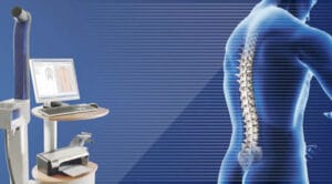
A female patient aged 67 with established osteoporosis, of which she had no knowledge, felt intense pain in the back, which continued for some time. An x-ray she did after 1 year showed a small sphenoid deformation in T9 and 66° kyphosis. Unfortunately, her physician suggested nothing and she continued without treatment.

After 2 years and because of intense pains she had another x-ray where further sphenoid deformation was ascertained in T9, but also sphenoid deformation in T8 and T10, and the kyphosis angle had increased to 84°.

The patient consulted another physician who gave her a treatment with bisphosphonates, but no support.
One and a half years later, it was ascertained by x-ray that the body of T9 had subdued to 20% of its total size and that T10 and T8 had further deteriorated. The angle had increased to 94°.

The picture was much worse by creating large thoracic hump.

The patient did not want to go ahead with kyphoplasty surgery and because the danger of deterioration was great, she was fitted with a special SPONDYLOS support brace.

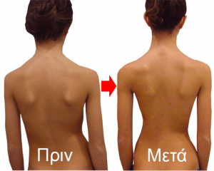
With the application of a brace and a better body posture, we have a reduction to elimination of pain in all cases.